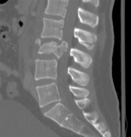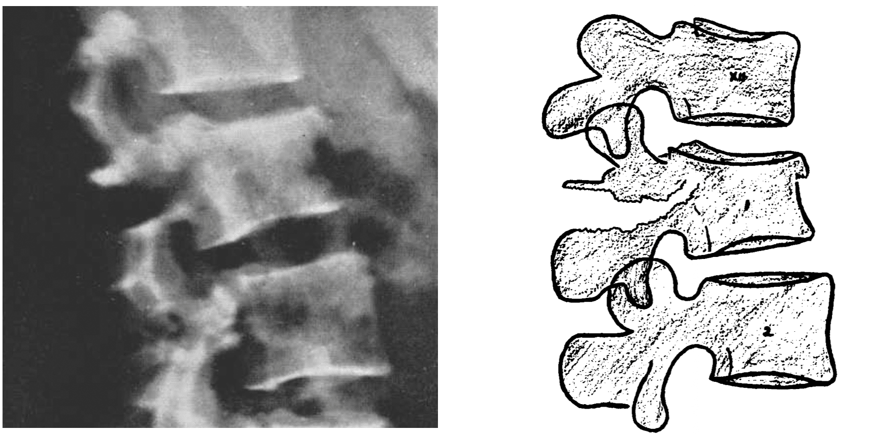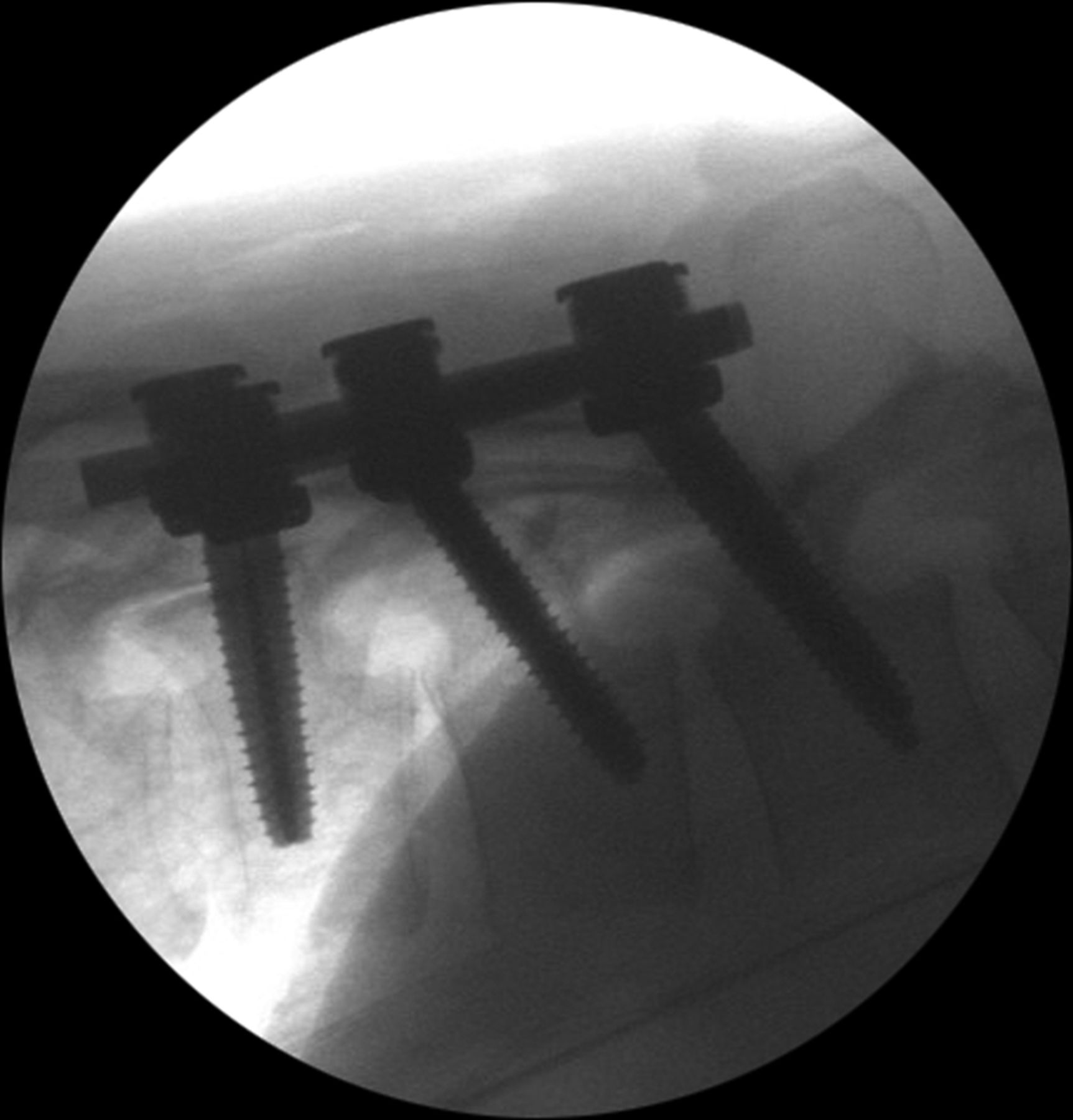

However, the ACR acknowledges in this document that initial use of CT or MRI of the thoracic and lumbar spine in this setting is controversial, but may be appropriate. The American College of Radiology lists radiography of the thoracolumbar spine as the most appropriate initial imaging for children less than 16 years of age with suspected thoracolumbar spine trauma. MRI findings of a Chance fracture may include a bright T2 signal (edema) surrounding low signal fracture lines, discontinuity of interspinous ligaments, injury to the intervertebral disc, spinal cord edema, and epidural hematoma. MRI should be performed in patients with clinical exam findings concerning spinal cord injury/compression or ligamentous injury. Magnetic resonance imaging (MRI) is superior to CT for the detection of soft tissue injuries, including injuries of the ligaments, spinal cord, cauda equina, and intervertebral discs. There may also be a distraction of the spinous processes and facet joints at the affected level. Obtaining coronal and sagittal reformations is critical to identifying a Chance fracture due to the horizontal orientation of the injury.Ĭommon CT findings of a Chance fracture include a horizontal fracture through the spinous process, pedicles, and vertebral body, with possible compression of the anterior vertebral body and buckling of the posterior vertebral body. If a trauma patient undergoes CT of the chest, abdomen, and pelvis, the included images of the spine are sufficient, and a separate dedicated spine CT is unnecessary.

Both the bladder and rectum should be examined.įor patients, 16 years of age or older with suspected thoracic or lumbar spine trauma, the most appropriate initial imaging study in the ACR Appropriateness Criteria is computed tomography (CT), and CT is superior to radiography for detection of fracture in this population. CT is also the imaging study of choice to assess for concurrent intra-abdominal injuries.

In addition, a thorough neurological exam is necessary. The “seat belt sign” can also be detected on computed tomography as stranding in the subcutaneous fat of the anterior abdominal wall.īecause of the potential for bowel and bladder rupture, it is highly recommended that a surgeon examine all trauma patients with abdominal pain. This finding is associated with Chance fractures and intra-abdominal injuries. Neurological deficits, such as weakness, numbness, and incontinence, can occur if there is a contusion of the spinal cord or cauda equina due to retropulsion of fracture fragments of the posterior vertebral body cortex.Ī significant physical exam finding to assess for is the “seatbelt sign,” which refers to bruising or abrasion on the abdomen in the distribution of a seat belt. Patients typically present with back pain without neurological deficits. In this scenario, the patient’s spine forcibly flexes over the lap seatbelt, which serves as a fulcrum. The most common mechanism of injury of a Chance fracture is a rapid deceleration in a motor vehicle collision. The most commonly associated intra-abdominal injuries are bowel perforations and mesenteric lacerations, both of which portend a high rate of patient mortality. The incidence of intra-abdominal injury can be as high as 50%. An osteoligamentous injury includes elements of both osseous and ligamentous injuries.Ĭhance fractures are unlike other spinal injuries because of their high association of intra-abdominal injuries. A ligamentous Chance injury can involve rupture of the interspinous ligament, posterior longitudinal ligament, ligamentum flavum, facet joint capsule, and intervertebral disc. An osseous Chance injury includes fractures of the spinous process, pedicles, and the vertebral body. A Chance fracture involves at least two columns–a distraction-type injury of the middle and posterior columns–with or without an additional compression-type injury of the anterior column.Ĭhance fractures can be further subdivided into osseous injuries, ligamentous injuries, and osteoligamentous injuries. Posterior column: posterior elements and posterior ligamentous complexĪn injury that involves at least two of the three columns is considered unstable.


 0 kommentar(er)
